
Figure 1.
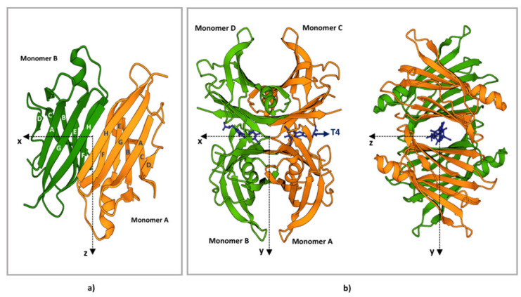
TTR structure. (a) Ribbon diagram of the AB dimer interface (b) Ribbon diagram of the TTR tetramers. The X-axis passes through the T4 binding channels, which are formed at the interface of monomers D-B and C-A. The halogen binding pockets (HBP) are shaped by the following amino acids: HBP1, Lys15-Leu17-Thr106-Val 121; HBP2, Lys15-Leu17-Ala108-Ala109-Leu110; HBP3, Ser117-Leu110-Thr119-Ala108. T4 is represented in blue. The figure has been produced using “www.rcsb.org”web site (protein ID code 1QAB), access date 22 June 2022.