Figure 3.
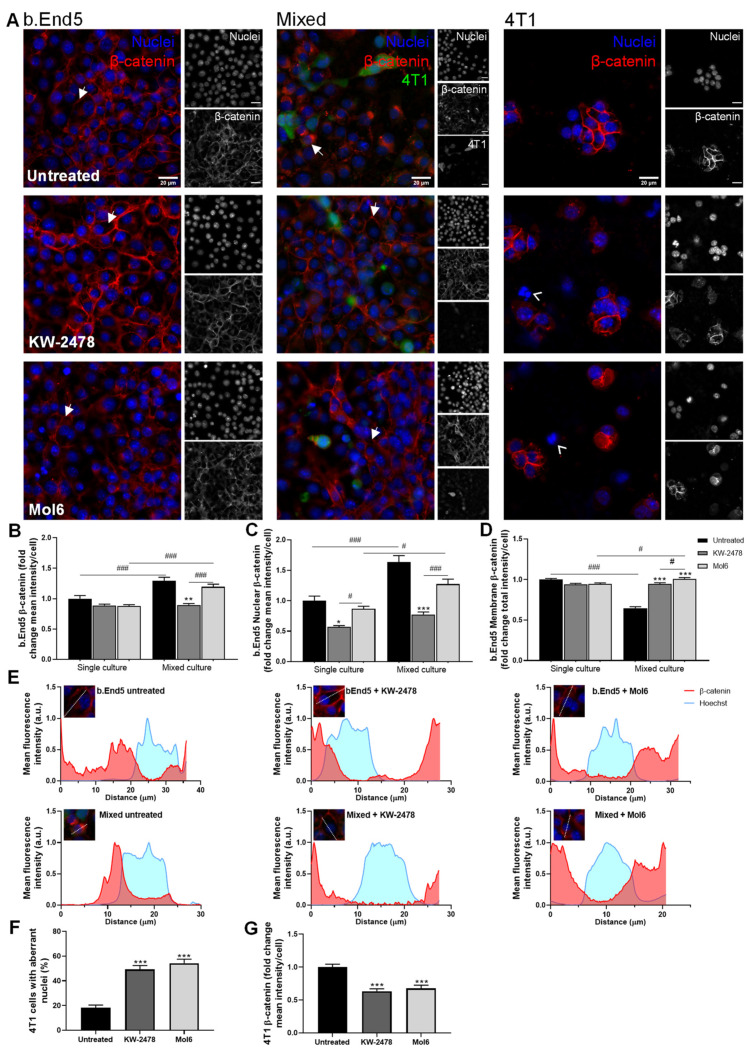
KW-2478 and Mol6 enhance barrier properties through increased membrane β-catenin expression in b.End5 cells exposed to 4T1 cells. Confluent monolayers of b.End5 cells under physiological laminar non-pulsatile shear stress were exposed to 4T1 cells (previously labelled with CellTracker™ CMFDA Green Dye,) and treated with KW-2478 or Mol6 (1 µM), or DMEM (untreated), for 24 h and the expression of the AJs protein β-catenin (red), in single and mixed cultures, was evaluated by immunofluorescence analysis. Hoechst 33342 was used as counterstaining for nuclei (blue). Scale bar: 20 µm. (A) Treatment with KW-2478 and Mol6 induced an increase in β-catenin expression in endothelial cells exposed to 4T1 cells, particularly at cell membrane (white arrows), and an increase in 4T1 cells with aberrant nuclei (white arrow heads). Semi-quantitative analysis of endothelial β-catenin (B) mean intensity per cell, (C) nuclear intensity, and (D) membrane intensity confirmed the observations. (E)The localisation of β-catenin was studied through plot profiles of pixel intensity throughout an endothelial cell (edge to edge). Semi-quantitative analysis of tumour cell cultures confirmed (F) an increase in the number of aberrant nuclei upon treatment with each drug, as well as (G) a decrease in β-catenin mean intensity per cell. Data are represented as mean ± SEM of three independent experiments, where 3 cells/field, 5 fields/condition were evaluated. Statistical significances are denoted as * p < 0.05, ** p < 0.01, and *** p < 0.001 vs. untreated conditions within the same culture type and # p < 0.05 and ### p < 0.001 between indicated groups.

