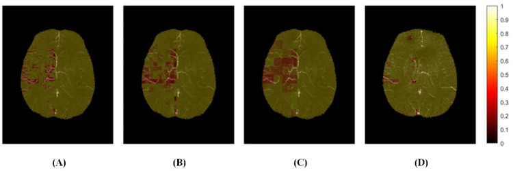
Figure 3.
The distribution map of cerebral venous oxygen saturation (SvO2) with different sizes of regions of interest (ROI) of a patient (40/72 slices). (A) ROI size was 3.59 × 3.59 × 1.6 mm3; (B,D) ROI size was 7.18 × 7.18 × 1.6 mm3; (C) ROI size was 14.36 × 14.36 × 1.6 mm3; (A–C): At admission; (D): At discharge.

