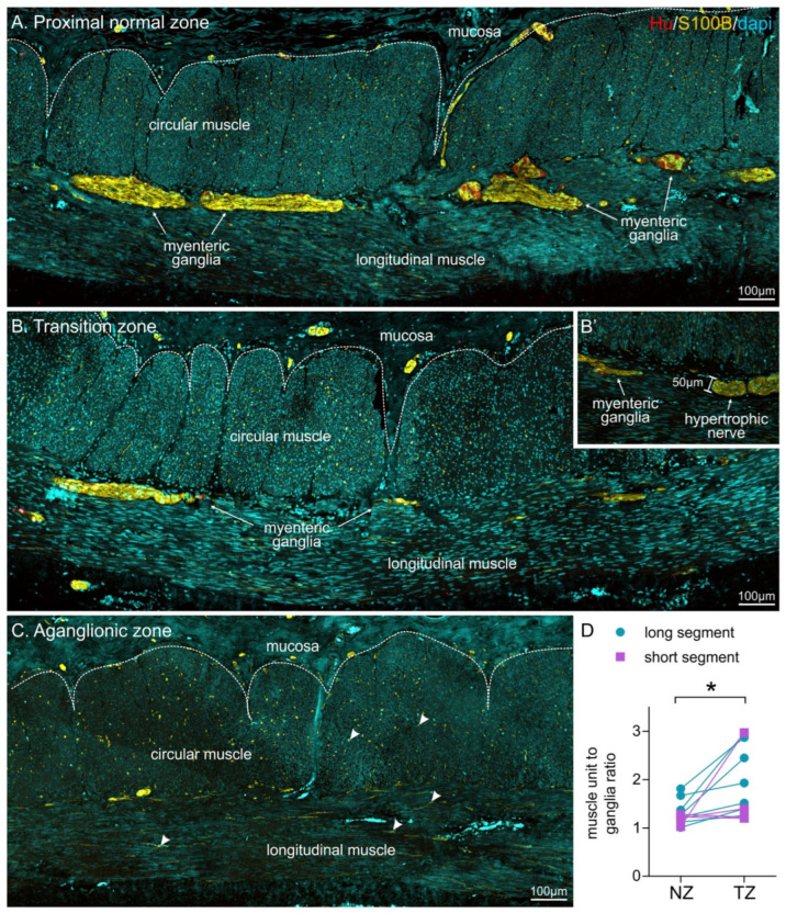
Figure 2.
Overview of the distribution of myenteric ganglia in relation to circular muscle units in the proximal normal zone (A, NZ), the transition zone (B, TZ), and the aganglionic zone (C) in longitudinal sections of HSCR patient tissue following immunohistochemistry against HuC/D (Hu), S100B and DAPI. Ganglia are more sparsely located in the transition zone, and no ganglia are visible in the aganglionic zone, although individual glial cells are present (some highlighted by arrowheads). (B’) shows a hypertrophic nerve bundle, which could be identified using S100B immunohistochemistry, but lacked neuronal cell bodies. Hypertrophic nerve bundles had a minimum width of 40 µm. (D) quantification of muscle unit to ganglion ratio in both long- and short-segment patients (* p = 0.012, n = 11, paired t-test).

