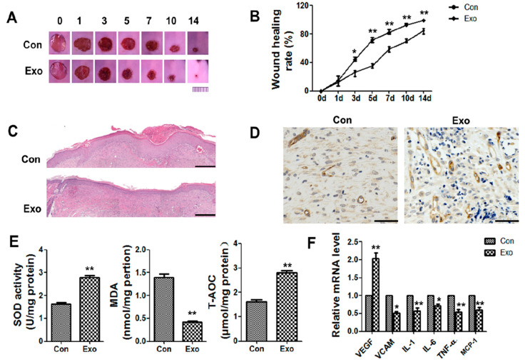
Figure 2.
Effects of ADSC-exos on wound healing in db/db model mice. (A) Digital images of wound areas treated with PBS or ADSC-exos on days 0, 1, 3, 5, 7, 10, and 14 (scale = 1 cm). (B) Analysis of the wound closure rate. (C) HE staining of normal skin or wound tissue treated with ADSC-exos on day 14 (scale = 500 μm). (D) CD34 staining of wound margins on day 14 (scale = 50 μm). (E) The levels of SOD, MDA, and T-AOC on day 7 were analyzed by SOD, lipid oxidation (MDA), and T-AOC detection kits. (F) mRNA levels of VEGF, VCAM, IL1, IL6, TNF-α, and MCP-1 on day 3 were analyzed by qRT-PCR. The data are shown as the mean ± standard deviation (SD; * p < 0.05, ** p < 0.01).

