Figure 3.
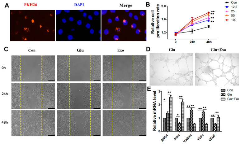
The effect of ADSC-exos on HUVECs. (A) Representative immunofluorescence images of PKH26-labelled exosomes in HUVECs. (B) The effect of ADSC-exo concentration on HUVECs activity was determined by CCK-8 assays. (C) The effect of ADSC-exos on HUVECs migration in a high-glucose environment was examined by scratch wound assays. The cells were divided into a normal control group, a high glucose control group, and an ADSC-exo-treated high glucose group. The images show the wound area in each group at 0 h, 24 h, and 48 h (scale = 250 μm). (D) Angiogenesis in different groups. The results showed that ADSC-exos enhanced angiogenesis in HUVECs. (E) mRNA levels of angiogenesis factors (ANG1, FILK1, and VEGF) and angiogenesis inhibitors (VASH1 and TSP1) were analyzed by qRT-PCR. The data are shown as the mean ± SD (* p < 0.05, ** p < 0.01).

