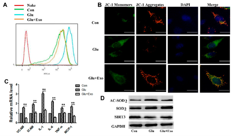
Figure 4.
ADSC-exos reduce ROS production in HUVECs in a high-glucose environment, increase the MMP, reduce inflammatory cytokine levels, and promote SIRT3 expression. (A) Analysis of HUVECs ROS levels by flow cytometry. (B) JC-1 signal in HUVECs was examined by fluorescence confocal microscopy. The cells were labeled with DAPI to show the nucleus (blue) and stained with JC-1 to show the mitochondria. Double staining of cells with JC-1 is shown: green for J-monomers, red for J-aggregates (scale = 50 μm). The data represent three independent measurements. (C) mRNA levels of VCAM, ICAM, IL1, IL6, TNFα, and MCP-1 in HUVECs were analyzed by qRT-PCR. (D) Western blot results showing the protein levels of AC-SOD2, SOD2, and SIRT3 in HUVECs treated with ADSC-exos in the high-glucose environment. The data are shown as the mean ± SD (** p < 0.01).

