. 2022 Jul 31;12(8):1850. doi: 10.3390/diagnostics12081850

Table 3.

The imaging configurations and main clinical distinctions of T1w, T2w, ceT1w, and FLAIR.

Sequence Sequence Characteristics Main Clinical Distinctions Example *
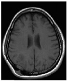
T1w Uses short TR and TE [64]
  • Lower signal for a higher water content [66], such as in edema, tumor, inflammation, infection, or chronic hemorrhage [66]

  • Higher signal for fat [66]

  • Higher signal for subacute hemorrhage [66]

graphic file with name diagnostics-12-01850-i001.jpg
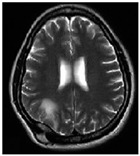
T2w Uses long TR and TE [64]
  • Higher signal for a higher water content, such as in edema, tumor, infarction, inflammation, infection, or subdural collection [66]

  • Lower signal for fat [66]

  • Lower signal for fibrous tissue [66]

graphic file with name diagnostics-12-01850-i002.jpg
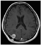
ceT1w Uses the same TR and TE as T1w; employs contrast agents [64]
  • Higher signal for areas of breakdown in the blood–brain barrier that indicate induced inflammation [65]

graphic file with name diagnostics-12-01850-i003.jpg
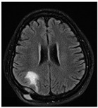
FLAIR Uses very long TR and TE; the inversion time nulls the signal from fluid [67]
  • Highest signal for abnormalities [65]

  • Highest signal for gray matter [67]

  • Lower signal for cerebrospinal fluid [67]

graphic file with name diagnostics-12-01850-i004.jpg

* Pictures from [68]. TR, repetition time. TE, echo time.