Figure 1.
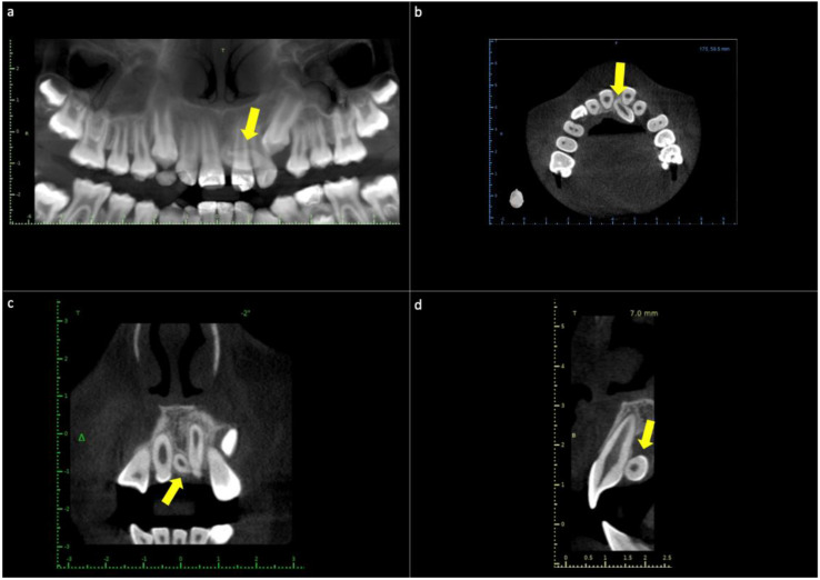
Cone−beam computed tomography (CBCT) of the maxilla. (a) The panoramic reconstruction shows the presence of a mesiodens supernumerary tooth between the midlines of maxillary central teeth. (b) Axial slice image showing the palatal location of the mesiodens in intimate relation to teeth 2.1 and 2.2. (c) Frontal slice image showing mesiodens between central incisors. (d) Sagittal slice image showing the palatal location of the supernumerary tooth in the crown-apical middle third of the central incisor. Yellow arrows—location of mesiodens.

