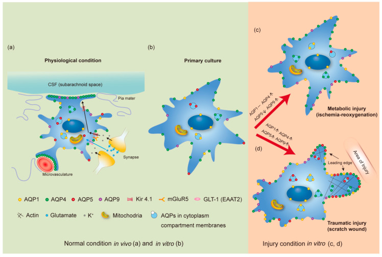
Figure 1.
AQP expression and subcellular distribution in astrocytes. The schematic representation summarizes the expression levels and subcellular localization of AQP1, AQP4, AQP5 and AQP9 in astrocytes under physiological conditions in vivo (a), in normal primary culture condition (b), and in primary culture upon metabolic (ischemia-reoxygenation) injury (c) and traumatic (scratch-wound) injury (d). Different colors are used to indicate the different subtypes: AQP1 (yellow), AQP4 (green), AQP5 (red) and AQP9 (purple). The symbol density reflects the comparative expression level of AQPs in specific regions, e.g., polarization of AQP4 to the astrocyte membranes facing CSF compartments or facing the microvasculature. In part (a), the directions of flow of K+ ions (black arrows) and glutamate molecules (blue arrows) in a tripartite synapse situation are indicated. The glutamate receptor (mGluR5), the glutamate transporter (GLT1/EAAT2), and the K+ transporter (Kir 4.1) known to interact with AQP4 are also depicted. Changes in the expression levels of AQPs between normal in vitro and the two injury conditions are indicated by bracketed symbols as increase [↑], decrease [↓], or unchanged [-]. Although these changes are shown only for in vitro conditions, similar changes for AQP4, AQP5 and AQP9 have been reported for in vivo conditions. Divergence for AQP1 expression change was demonstrated (not shown in the figure). Stable levels of AQP1 were determined by us in primary culture of mouse astrocytes after ischemia treatment and mouse MCAO model [53,131]. However, the other two studies reported induction of AQP1 expression in astrocytes using the rat MCAO model and human ischemic brain lesions when compared to physiological conditions [132,133].

