
Figure 1.
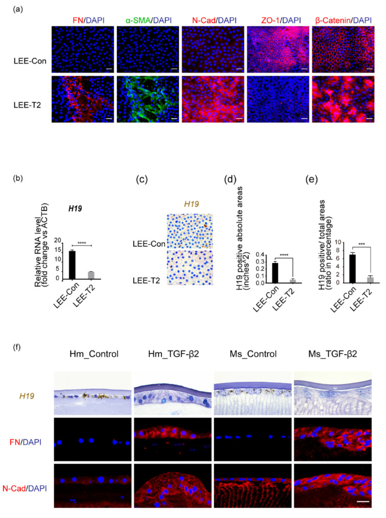
LncRNA H19 was highly expressed in normal lens epithelium while downregulated by exposure to TGF-β2. (a) Immunofluorescent staining of mesenchymal markers (FN, α-SMA, and N-Cad), and epithelial markers (ZO-1, β-catenin), on human lens epithelial explants (LEEs), when exposed to TGF-β2 (10 ng/mL, 48 h; LEE-T2), in comparison to control group (LEE-Con). Scale bars, 20 μm. (b) RNA analysis of LEEs-Con and LEEs-T2 (age-matched, n = 6) was performed using β-actin as the internal control. (c) H19 probes were hybridized in situ. Scale bars, 20 μm. (d,e) Image J was used to quantify the absolute value of H19 positive areas (d) and the ratio of H19 positive to total area within each cell (e). At least three experiments were repeated, and data were shown as mean ± SD. *** p < 0.001, **** p < 0.0001 vs. LEE-Con. (f) RNAscope targeting H19 on the freshly cut sections from human and mouse whole lenses was performed. The lower two panels showed immunostaining of mesenchymal markers FN and N-Cad. Scale bars, 20 μm.