
Figure 2.
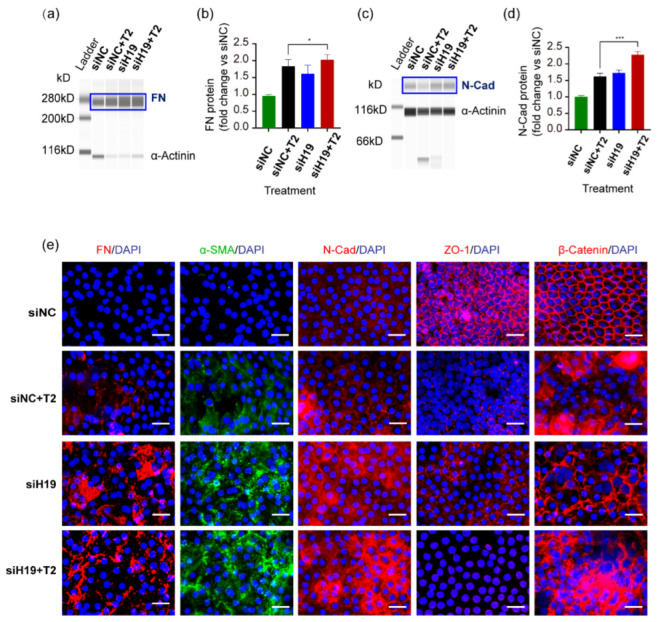
Knockdown of H19 in human lens epithelial cells in situ accelerated TGF-β2-induced EMT. (a–d) After transfection with siH19-002 for 24 h and further exposure to TGF-β2 for 24 h, the Wes platform detected protein levels of mesenchymal markers FN and N-Cad in the human LEEs, which were further quantified using Image J. * p < 0.05, *** p < 0.001. (e) Fluorescent staining of FN, α-SMA, N-Cad, ZO-1, and β-catenin on human LEEs. Scale bars, 20 μm.