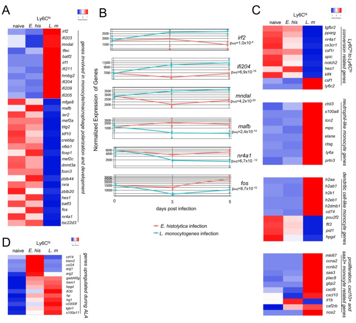
Figure 3.
Early M2 polarization and a less activated mRNA expression profile in Ly6Chi monocytes from E. histolytica-infected compared to L. monocytogenes-infected mice. (A) Heat map showing selected regulated genes (padj < 0.05; foldchange > 2) involved in monocyte/macrophage polarization and activation of Ly6Chi monocytes derived from the livers of E. histolytica (E. his)- and L. monocytogenes (L. m)-infected. (B) Time-course analysis of mRNA encoding M2 transcription factors and interferon-regulated/activated factors. (C) Heatmap showing classification of Ly6Chi monocytes derived from the livers of both infection models according to expression of genes involved in conversion from Ly6Chi to Ly6Clo, neutrophil-like, dendritic cell-like, or Cxcl10+ and Saa3+-like monocytes. (D) Heat map of selected genes upregulated during ALA. All heatmaps were designed using the online tool “heatmapper” [34].

