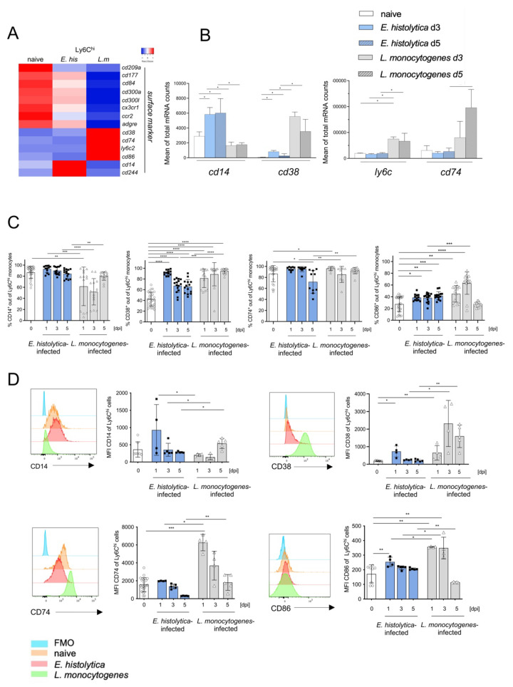
Figure 4.
Marked differences in expression of surface markers by Ly6Chi monocytes after infection with E. histolytica or L. monocytogenes (A) Heatmap depicting differential expression of mRNA encoding selected markers on the surface of liver Ly6Chi monocytes after infection with E. histolytica (E. his) or L. monocytogenes (L. m). (B) Normalized mRNA counts of selected surface marker genes (from transcriptome analysis). (C) Percentage of CD14+, CD38+, CD74+, and CD86+ Ly6Chi monocytes during the course of infection at the indicated time points post-infection (measured by flow cytometry). (D) Histogram and MFI of CD14+, CD38+, CD74+, and CD86+ Ly6Chi monocytes during the course of infection. Data in C were pooled from three independent experiments. Data in D are representative of one of these three experiments and all data are presented as the mean ± SEM (* p < 0.05; ** p < 0.01; *** p < 0.001, **** p < 0.0001; Mann-Whitney U test).

