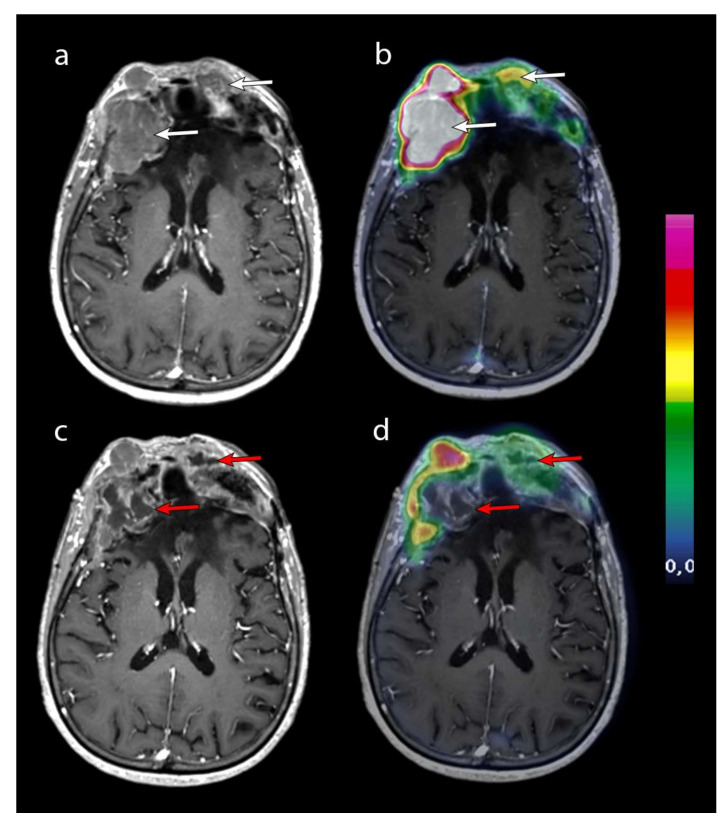
Figure 2.
Contrast-enhanced T1-weighted axial MRI after the second cycle of PRRT (a) and after the fourth cycle of PRRT (c) was merged with 68GA-DOTATOC PET (respectively, panels (b) and (d)). Two frontal lesions (white arrows) with high 68GA-DOTATOC uptake necrotized after 4 cycles (red arrows). The question remains as to the origin of this necrosis, which may be a direct effect of PRRT or a natural necrotic tumor progression. Patient 2 was the only patient to progress with this necrotic appearance. Unequivocal progression of the other lesions was assessed according to RANO criteria on the MRI performed 4 months later.

