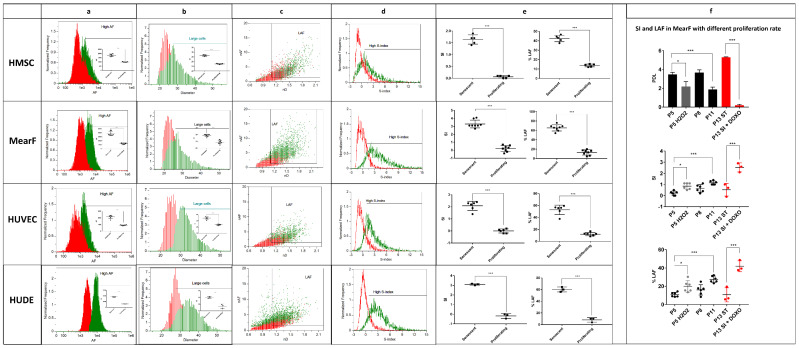
Figure 2.
Quantitative estimation of the senescent index (SI) and % of large autofluorescent cells (LAF) in various senescence models. (a) Overlayed histograms and quantitative estimation of autofluorescence (AF) in proliferating (red) and senescent cells (green). All senescent samples display increased autofluorescence compared to non-proliferating samples. (b) Overlayed histograms and quantitative estimation of the diameter (D = (width + height)/2)) in proliferating (red) and senescent cells (green). All senescent samples display increased diameter compared to non-proliferating samples. (c) Overlayed representative dot plots of normalized autofluorescence (nAF = “AF”/“mean AF of proliferating samples”) vs. normalized diameter (nD = “D”/“mean D of proliferating samples”) showing that LAF increases in senescent samples (green events) compared to non-senescent samples (red events). The threshold of nAF was set at 1.5 whereas the threshold of D was set at 1.1 for all samples. (d) Overlayed representative histograms of the SI (SI = ((nAF − 1) + 5 × (nD − 1))/2) in proliferating (red) and senescent cells (green). (e) Quantitative estimation of SI and LAF in proliferating and senescent HMSC (n = 5), MearF (n = 8), HUVEC (n = 6) and HuDe (n = 3). (f) Comparison of SI and LAF in MearF with different population doubling level (PDL). SI and LAF significantly increase after stressing conditions that decrease PDL, such as treatment with H2O2 (250µM H2O2 × 2 h + 1 week of resting, n = 6) or at late passages (P11, n = 6) compared to early passages (P5, n = 6). Spontaneous transformation of one of the cultures (P13 ST, n = 3 replicates from the same culture) resulted in a strong increase in PDL and a parallel decrease in SI and LAF. Treatment of P13 ST with doxorubicin 75 nM × 1 week (P13 ST + DOXO, n = 3 replicates from the same culture) strongly increased both SI and LAF. MearF = mouse ear fibroblasts; HUVEC = human umbilical vein endothelial cells; HMSC = human bone marrow mesenchymal stem cells; HuDe = human dermal fibroblasts; * p < 0.05; *** p < 0.001 by Student’s t test.

