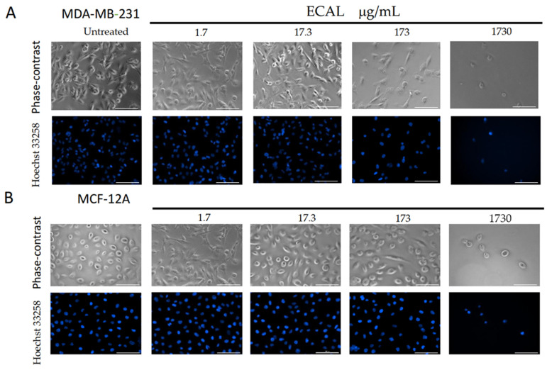
Figure 2.
Morphological changes induced by increasing concentrations of the hydroethanolic extract of C. album leaves (ECAL) (1.7–1730 μg/mL) incubated for 24 h in (A) MDA-MB-231 cells or (B) MCF-12A cells. Upper panel—representative images from five independent experiments obtained under an objective lens of a phase-contrast of the Lionheart microscope; lower panel—representative images from five independent experiments with nuclei stained with Hoechst 33258 (blue) obtained under an objective lens of a Lionheart microscope. Scale bar = 100 μM.

