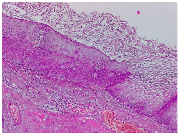
Figure 9.
Histological specimen of the previous case illustrating CIN 3 lesion—proliferation of squamous cells with abnormal mitosis throughout the entire thickness and acanthosis and parakeratosis of the superficial layers. Right: Koilocyte transformation illustrating the simultaneous presence of two lesions of different severity levels. (Hematoxylin–eosin 10×).

