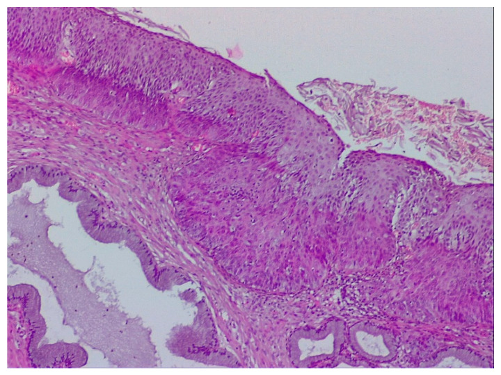
Figure 11.
Image showing three typical aspects of CIN in the case in Figure 8: squamous abnormalities and glandular dilatations as well as moderate ulceration of the squamo–cylindric junction with anisocoria and nuclear polymorphism. (Hematoxylin–eosin 10×).

