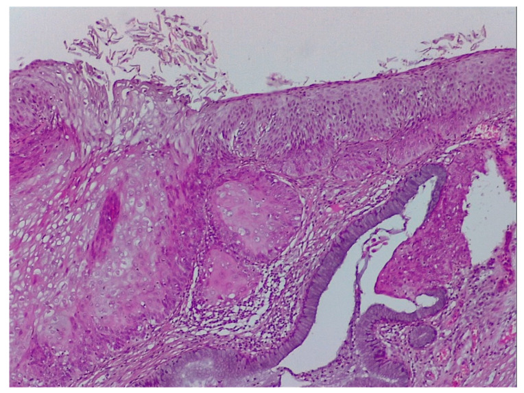
Figure 13.
Pathological specimen of the case described in Figure 9: microinvasive carcinoma, superficial ulceration, broken basal membrane, and presence of the keratinization foci in stroma. The columnar epithelium is normal. (Hematoxylin–eosin 10×).

