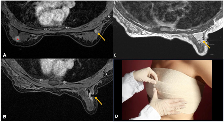
Figure 2.
(A). Staging MRI in a woman with known left breast cancer (red asterisk) showed focal non-mass enhancement in the right breast (yellow arrow). (B). MRI-guided biopsy to rule out contralateral synchronous cancer showed the tip of the obturator at the right location (yellow arrow). (C). Post-biopsy non-fat saturated image showed a large hematoma at the biopsy site almost occupying the upper outer quadrant (yellow arrow). (D). Tight compression bandage applied to the breast after the biopsy reduces the risk of further bleeding significantly.

