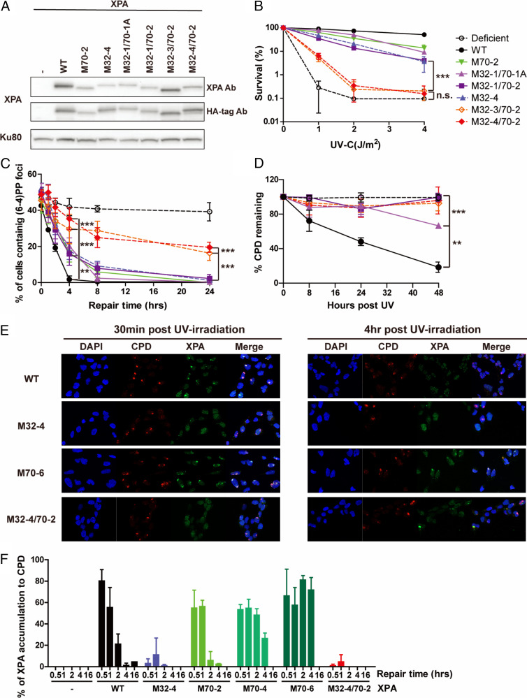
Fig. 4.
The RPA32 and 70 binding domains of XPA synergistically contribute to NER activity in cells. (A) XPA expression levels in XP2OS cells transduced with WT and RPA32/70-interaction mutant XPA detected anti-XPA and anti-HA antibodies, using Ku80 as a loading control. (B) Clonogenic survival assays. Cells were treated with the indicated UV dose, grown for 10 d and stained with methylene blue. Survival rates were normalized to nontreated cells. The P value was compared to M32-4. (C) Quantification of (6, 4) PPs repair at sites of local UV damage. Cells were irradiated through a 5-µM micropore filter with UV (100 J/m2) and stained for (6, 4) PP. One-hundred cells were counted for each sample and the data represent at least two independent experiments. The P value was compared to XPA WT. (D) Determination of CPD repair kinetics using slot-blot assays. Cells were irradiated with 5 J/m2 genomic DNA isolated at the indicated time points and adduct levels determined with an anti-CPD antibody. Band intensities were normalized to WT at 0 h. The P value was compared to XPA WT. (E) Representative figure of colocalization assay with CPD and XPA. Cells were irradiated through a 5-µM micropore filter with UV (100 J/m2) and stained for CPD and XPA to measure the accumulation of XPA to CPD. XPA was stained by HA-tag antibody. For microscope analysis, 10X magnification was used with Axio observer 7(F) Quantification of E: 100 cells were counted for each sample and data represent two independent experiments. Percent of colocalization was calculated by dividing number of cells containing colocalization by number of cells containing CPD foci. **P < 0.01, ***P < 0.001.

