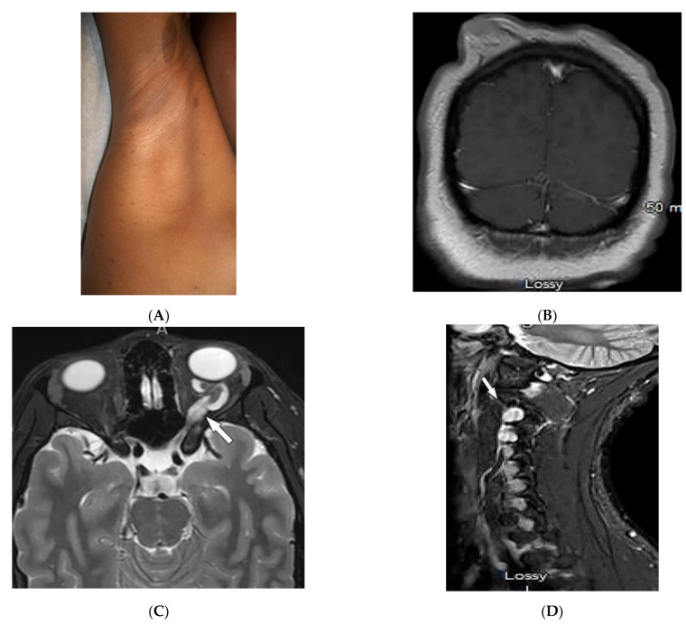
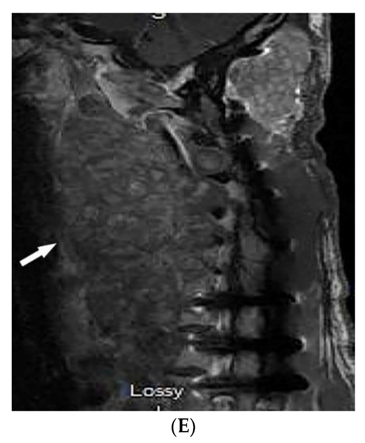
Figure 9.
Dermatologic and radiologic images representative of neurofibromatosis type 1 (NF1): (A) Café-au-lait macules on the upper arm and multiple small macules on the axillae (axillary “freckling”). (B) Coronal post-contrast T1WI of the brain demonstrates diffuse cutaneous neurofibromatosis. (C) Axial T2WI of the brain shows diffuse thickening of the left optic nerve (arrow), consistent with optic glioma. (D) Sagittal T2WI of cervical spine demonstrates intraneural foraminal neurofibromas (arrow). (E) Sagittal T1WI of cervical spine in a 56-year-old male demonstrates plexiform neurofibroma (arrow).


