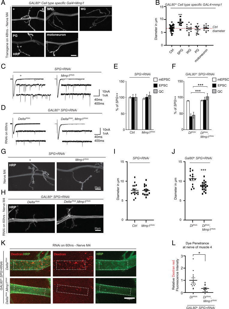
Fig. 5.
Mmp1 in SPG is responsible for defects associated with loss of Delta. (A) Maximum-intensity projection of a confocal Z stack of the larval motor nerve at the NMJ of A3M4 of w1118 (control), moody-GAL4 (SPG), nrv2-GAL4 (WG), c527-GAL4 (PG), and BG380-GAL4 (motoneuron) crossed to tub-Gal80ts;UAS-Mmp1 transferred to 27 °C for 48 h and stained with anti-HRP. Scale bar = 20 micron. (B) Diameter of the nerve as delineated by HRP for the genotypes in A, n = 33 for control, n = 10 for SPG, n = 12 for WG, n = 12 for PG, and n = 12 for motoneuron, followed by one-way ANOVA and Tukey’s test for multiple comparisons. (C) Representative traces of mEPSCs and EPSCs from moody-GAL4 crossed to w1118 (control) or UAS-Mmp1RNAi. (D) Representative traces of mEPSCs and EPSCs from tub-GAL80ts/moody-GAL4;UAS-DeltaRNAi or tub-GAL80ts/moody-GAL4;UAS-DeltaRNAi/UAS-Mmp1RNAi transferred to 29 °C for 60 h. (E) Quantification of mEPSCs, EPSCs, and QC from genotypes described in C, n = 15 for control and n = 13 for UAS-Mmp1RNAi followed by Student’s t test for each respective pair. (F) Quantification of mEPSCs, EPSCs, and QC from D, DeltaRNAi (n = 15) or DeltaRNAi, Mmp1RNAi (n = 17) followed by Student’s t test for each respective pair. (G) Maximum-intensity projection of a confocal Z stack at the larval motor nerve of A3M4 of the NMJ of moody-GAL4 crossed to w1118 (control) or UAS-Mmp1RNAi stained with anti-HRP. (H) Maximum-intensity projection of a confocal Z stack at the larval motor nerve at the NMJ of A3M4 of tubGAL80ts/moody-GAL4;UAS-DeltaRNAi or tubGAL80ts/moody-GAL4;UAS-DeltaRNAi/UAS-Mmp1RNAi transferred to 29 °C for 60 h and stained with anti-HRP. (I) Diameter of the nerve as delineated by HRP for the corresponding genotypes in G, n = 16 for control and n = 14 for Mmp1RNAi, followed by Student’s t test. (J) Diameter of the nerve as delineated by HRP for the corresponding genotypes in H, n = 13 DeltaRNAi and n = 18 for DeltaRNAi, Mmp1RNAi, followed by Student’s t test. (K) Maximum-intensity projection of a confocal Z stack at the larval motor nerve at the NMJ of A3M4 of tubGAL80ts/moody-GAL4;UAS-DeltaRNAi or tubGAL80ts/moody-GAL4;UAS-DeltaRNAi/UAS-Mmp1RNAi transferred to 29 °C for 60 h followed by injection with dextran (red) and stained with anti-HRP (green). The dotted areas show the area chosen to assess dye intensity. (L) Quantification of the relative dextran fluorescence intensity signal in K, n = 12 for DeltaRNAi and n = 10 for DeltaRNAi, Mmp1RNAi followed by Student’s t test. *P < 0.05; **P < 0.01 and ***P < 0.001. All error bars are standard error of the mean.

