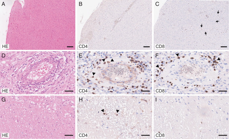
Fig. 1.
Localization of CD4+ T cells and CD8+ T cells in the GBM site. Histologic analysis of GBM tissue (overview; A–C) comparing perivascular (D–F) and necrotic areas (G–I). Hematoxylin and eosin (HE) staining (A, D, and G) and immunohistologic staining for CD4 (B, E, and H) and CD8 (C, F, and I). (Scale bars, 200 μm [A–C] and 50 μm [D–I].) Note that CD4+ T cells were present in the vessel walls (arrowheads in E) and in necrotic tissue (arrowheads in H) while CD8+ T cells were largely localized in perivascular areas (arrowheads in F) or scattered in GBM tissue (arrows in C).

