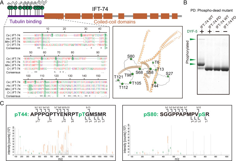
Fig. 1.
DYF-5 phosphorylates the tubulin-binding region of IFT-74. (A) Schematic diagrams (Top), homologous sequence alignment (Bottom Left), and cartoon structural representation (Bottom Right) showing the identified phosphorylation sites in C. elegans IFT-74. The phosphorylation residues and their possible corresponding homologous sites were labeled with green shadows in the alignment. The structural coordinate of IFT-74 from AlphaFold Protein Structure Database (ID: A0A2C9C2L6) was used to generate the structural figure. (B) Phos-tag kinase assays validating the determined phosphorylation sites. Purified WT and phospho-dead (PD; Ser-Ala/Thr-Ala) mutant IFT-74 (1-372) protein was treated with DYF-5 kinase and evaluated by Phos-tag SDS-PAGE. The phosphorylation-induced bandshifts are indicated by green arrowheads. (C) Representative LC-MS/MS results of the determination of IFT-74 phosphorylation sites.

