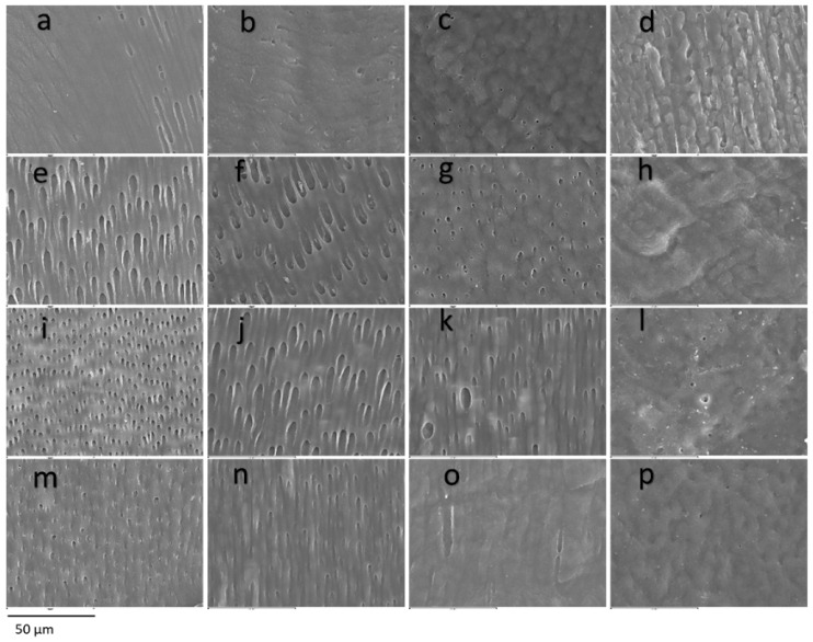
Figure 3.
Dentin. SEM photomicrographs at original magnification of 1000×. Superficial dentin (a–d) of a control tooth (a), dentin with mild involvement and alteration in the direction of the dentin tubules (b), dentin with moderate involvement, heterogeneity in the diameter and direction of the dentin tubules and cerebroid appearance (c) and severely affected dentin, atubular amorphous tissue with a cerebroid appearance (d). Middle dentin (e–h) of a control tooth (e) without affectation, with mild affectation (f) with heterogeneity in the diameters of the dentinal tubules, moderate with partial obliteration of the dentinal tubules (g) and severe affectation with the presence of amorphous atubular tissue (h). Deep dentin (i–l) of a control tooth (i) with homogeneity in direction and tubular diameter, mildly affected dentin (j) presenting slightly irregular diameters, moderately affected dentin (k) with the presence of giant tubules and heterogeneity in the diameter of the dentinal tubules and severely affected dentin (l) practically atubular and with an amorphous appearance. Pulpar dentin (m–p) of a control tooth (m) without alterations, of a tooth with slight alterations (n) with slight discrepancies in the tubular diameter, of a tooth with moderate alterations (o) with a decrease in the tubular pattern and of a tooth with severe alterations (p) with an atubular and cerebroid pattern.

