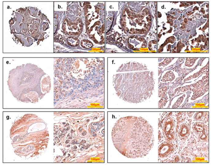
Figure 7.
Histology of ACBD3 stained breast cores. (a–d) An invasive carcinoma from a 39-year-old female, stage IIA, ER- PR- HER2 2+ reveals a pattern of differential ACBD3 staining (brown), haematoxylin was used a as a nuclear stain (blue). (a) Low magnification image of entire core, fibrous interlobular tissue has a low level of ACBD3 staining whilst breast duct acini have high levels of ACBD3 staining. (b) Medium magnification of regular and irregular duct acini. (c) High magnification of regular small acini, shows high ACBD3 staining of luminal epithelial and basal myoepithelial cells. (d) High magnification of irregular large acini. Luminal epithelial and basal myoepithelial cells are both highly stained for ACBD3 but cells within the acini, possibly a ductal carcinoma in situ have a moderate-to-low level of ACBD3 staining, with some embedded cells with high levels of ACBD3, possibly luminal epithelial cells. (e–h) Typical ACBD3 staining of different cores. (a) Invasive ductal carcinoma of a 47-year-old female. Stage IA, grade 3, T1N0M0 scored as 1.1 overall for ACBD3 staining. (b) Invasive ductal carcinoma of a 50-year-old female. Stage IIA, Grade 2, T2N0M0 scored as a 2 overall for ACBD3 staining. (c) Normal-adjacent breast tissue with ductal ectasia of a 41-year-old female, scored 2.9 overall. (d) Cancer-adjacent tissue (adenosis) of a 39-year-old female, scored 1.7 overall.

