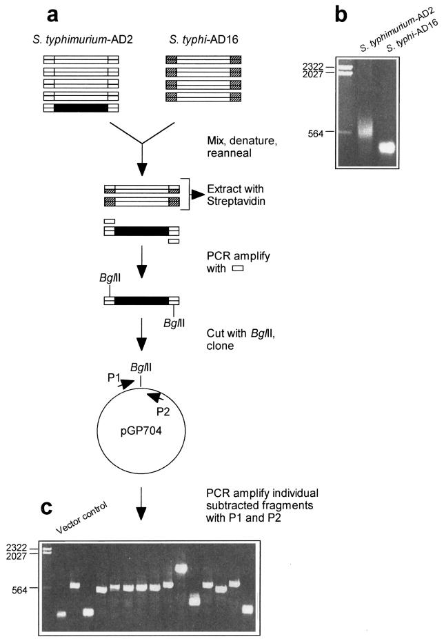
FIG. 1.
Genomic subtraction procedure used to isolate S. typhimurium-specific DNA fragments. (a) Schematic representation of the subtraction procedure. A fragment of S. typhimurium DNA not present in S. typhi is shown in black. Biotinylated adapter sequences are indicated by hatching. (b) Agarose gel (sizes [in kilobases] of marker fragments are indicated) showing PCR-amplified S. typhimurium-AD2 fragments and S. typhi-AD16 fragments before subtraction. (c) Agarose gel showing individually amplified S. typhimurium DNA fragments after subtraction and cloning. The PCR product obtained by using the cloning vector without an insertion as the template is in the lane adjacent to the size marker lane. (See text for further details.)

