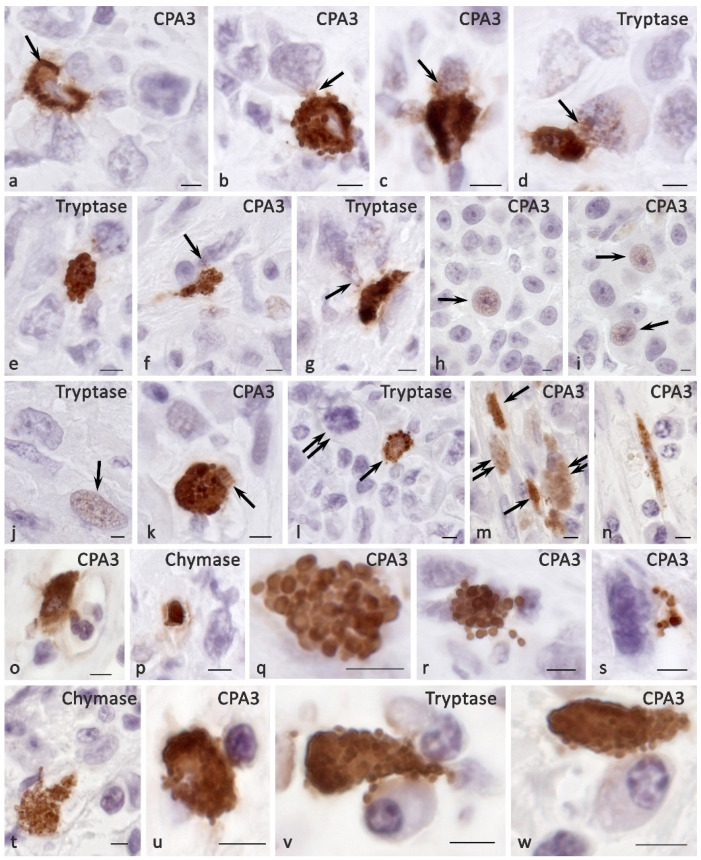
Figure 5.
Histotopography and secretory pathways of specific MC proteases in the tumor microenvironment. (a) Transgranulation of CPA3 simultaneously towards several tumor cells. (b) Secretion of CPA3 in the composition of the granules to the tumor cell. (c–g) Variants of MC-specific protease secretion with signs of transfer into the nucleoplasm of tumor cells (indicated by an arrow). (h–j) Nuclei of tumor cells immunopositive to CPA3 (h,i) and tryptase (j) in the MC absence in the zone of paracrine effects (indicated by an arrow). (k) Massive secretion of CPA3 into the locus of the tumor microenvironment (indicated by an arrow). (l,m) Colocalization of MCs (indicated by an arrow) with tumor cells (indicated by a double arrow) within paracrine effects. (n,o) Adjacence of MCs with elements of the microvasculature. (p–s) Various options of the secretory activity of protease-containing fragments of the MC cytoplasm (p,q) and individual secretory granules (r,s). (t) Adjacence of MC with fibroblast. (u–w) Targeted secretion of specific MC proteases towards the plasmalemma of a lymphocyte (u) and plasmocytes (v,w). Scale bar: 5 µm.

