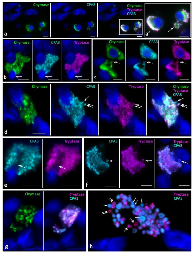
Figure 6.
Protease phenotype and sectoral activity of MCs during the formation of macrovesicles and cytoplasts in the tumor microenvironment. (a) Eccentric position of the nucleus in MCs with the expression of a triad of specific proteases, with signs of detachment of a large cytoplasm fragment from one of them (indicated by an arrow). (b) Signs of the start of the nucleus release and MC enucleation (indicated by an arrow). (c) Colocalization of protease-positive MCs with a nuclear-free backbone of the cytoplasm with signs of secretory activity (indicated by an arrow). (d) Enucleation of the nucleus is accompanied by selective secretion as part of CPA3 granules (indicated by an arrow), or together with tryptase (indicated by a double arrow). (e) Completion of the nucleus exit from the MC with the formation of a cavity in the cytoplasm (indicated by an arrow). (f–h) The terminal stage of MC enucleation with the formation of a cytoplasmic backbone and preservation of the stock of specific proteases in the composition of the granules and secretory activity (f), indicated by an arrow). (g,h) Splitting the mother cytoplasm into two smaller fragments, the granules have a variability in the content of specific proteases, including granules with predominant accumulation of CPA3 (indicated by an asterisk) and tryptase (indicated by a double arrow). Scale bar: 5 µm.

