Figure 7.
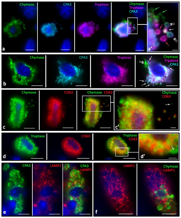
Morphological equivalents of secretory pathways of specific MC proteases in the tumor microenvironment. (a) Different phenotype of specific proteases of secretory granules in the intercellular substance after secretion: tryptase+ (red arrow), chymase+ (green arrow), tipase+chymase+ (white arrow), chymase+tryptase+CPA3+ (double arrow). (a’) The same as (a) at higher magnification. (b) Simultaneous secretion of tryptase combined with CPA3 (indicated by an arrow), and chymase (indicated by a double arrow). (c,d) High degree of immunopositivity to CD63 of chymase-containing and tryptase-containing MCs. Secretory granules differ in the degree of immunopositivity for CD63 in the extracellular matrix (c’) and MC cytoplasm (d’), including the absence (indicated by the arrow). (e,f) LAMP1 expression in CPA3+ mast cell (e) and chymase+ MCs (f).

