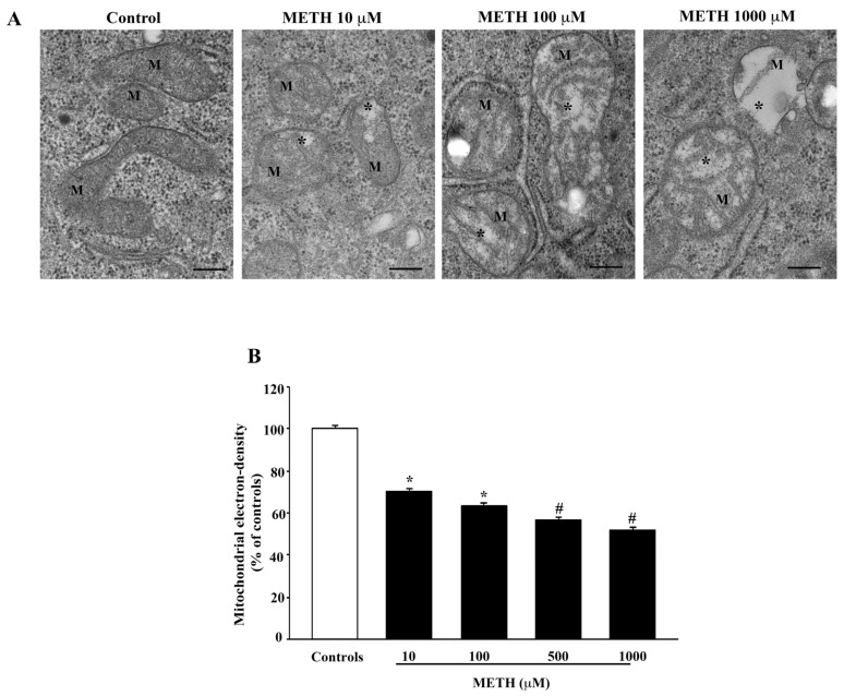
Figure 8.
METH decreases matrix electron-density. (A) Representative TEM micrographs showing a dose-dependent decrease in electron density of mitochondrial matrix. In control, mitochondria possess a marked electron dense matrix, whereas after increasing doses of METH (from 10 μM up to 1000 μM) the mitochondria possess a diluted matrix that appears less electron dense compared with control (*). M = mitochondria. (B) Graph reports values showing matrix dilution as a weighted measurement (percentage of matrix electron density from controls). Values are given as the percentage mean ± S.E.M from N = 150 mitochondria per group. * p ≤ 0.05 compared with controls; # p ≤ 0.05 compared with controls and METH 10 μM and 100 μM. Scale bars = 160 nm.

