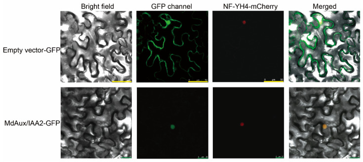
Figure 3.
Subcellular localization of MdAux/IAA2. NF-YA4-mCherry was the nuclear marker. MdAux/IAA2-GFP was MdAux/IAA2, which was connected to the carrier of PRI101 containing the GFP label. The empty vector-GFP was the control. The empty vector-GFP yellow scale was 50 μm, and the MdAux/IAA2-GFP green scale was 25 μm.

