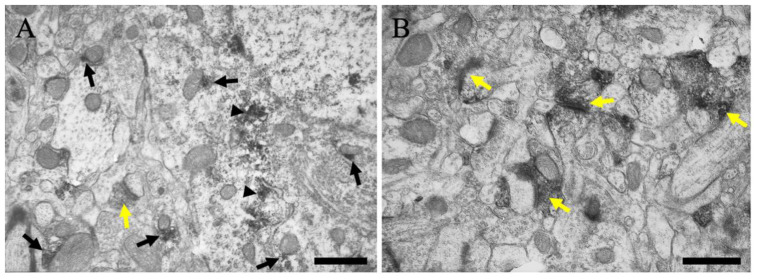
Figure 5.
Immunoelectron microscopic views of layer V of the cerebral cortex are shown. (A) The neuron and neuropil near the neuronal soma are presented. (B) The neuropil far from the neuronal soma is presented. Black arrows indicate MGRN1 immunoreactivity near the mitochondria. Black arrowheads indicate MGRN1 immunoreactivity near the subcellular organelles without mitochondria; e.g., endoplasmic reticulum. Yellow arrows indicate MGRN1 immunoreactivity in the pre-synapses. Scale bars = 500 μm.

