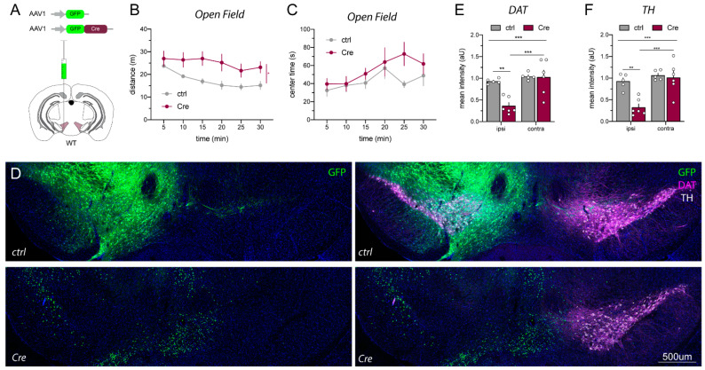
Figure 4.
DAT and TH loss in the VTA of AAV1_Cre-GFP-injected WT mice coincide with hyperactivity. (A) Scheme visualizing unilateral injection of Cre (AAV1_Cre-GFP) and AAV1_GFP controls at 1013 GC/mL into the VTA of adult WT C57BL/6J mice (for details see Figure S1). (B,C) Cre-injected WT mice become hyperactive compared to GFP control-injected mice in the open field (ctrl n = 5, Cre n = 6; mixed-effect analysis, distance: F(2.327,20.47) = 8.179, p = 0.4653 and genotype: p = 0.0465 *, center time: F(5,45) = 1.380, p = 0.2498 and genotype: p = 0.2969). (D) Immunostainings of unilaterally injected WT mice with either AAV1_GFP (ctrl; top panels) and AAV1_Cre-GFP (Cre; bottom panels) four weeks after injection. (E,F) Quantification revealed a massive loss of TH (white) and DAT (magenta) in the ipsilateral (ipsi, injected) side of Cre-injected mice compared to the contralateral (contra, non-injected) side and AAV1_GFP control-injected mice (ctrl n = 5, Cre n = 6; two-way ANOVA, DAT: F(1,9) = 12.12, p = 0.0006 ***; ipsi ctrl vs. Cre p = 0.0019 **; Cre ipsi vs. contra p = 0.0003 ***; TH: F(1,9) = 9.171, p = 0.0143 ***; ipsi ctrl vs. Cre p = 0.0011 **; Cre ipsi vs. contra p = 0.0006 ***). Scale bar 500 µm.

