Figure 2.
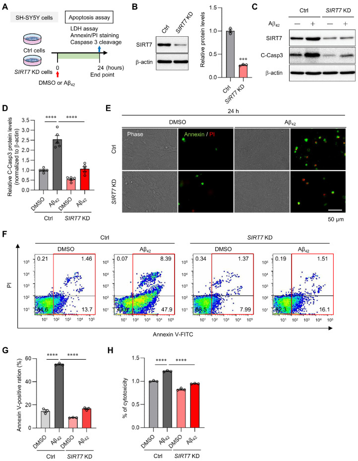
SIRT7 KD improves Aβ-induced apoptosis. (A) Experimental scheme for analyzing Aβ42-induced apoptosis in SIRT7 KD SH-SY5Y cells. (B) SIRT7 KD efficiency was confirmed by Western blot analysis when SH-SY5Y cells were transfected with SIRT7 siRNA for 48 h. (C) After SH-SY5Y cells had been transfected with control and SIRT7 siRNA for 48 h, cells were treated with 5 μM Aβ42 for 24 h. Western blot analysis of cleaved caspase 3 was performed. (D) The value of cleaved caspase 3 was normalized to that of β-actin. (E) Representative microscopy images of fluorescent annexin V (green)- and PI (red)-stained cells are shown for cells treated in the same condition as that in Figure 2A. Scale bar, 50 μm. (F) Flow cytometry analysis was performed on cells treated in the same condition as that in Figure 2A. Representative flow cytometry plots using annexin V-FITC/PI staining for apoptosis. (G) Percentage of total annexin V-positive cells was calculated. (H) Cell death was evaluated by an LDH activity assay on cells treated in the same condition as in Figure 2A. All data are shown as the mean ± SEM. Statistical significance was determined by either Student’s t-test or two-way ANOVA with Tukey’s post hoc test. *** p < 0.001; **** p < 0.0001.

