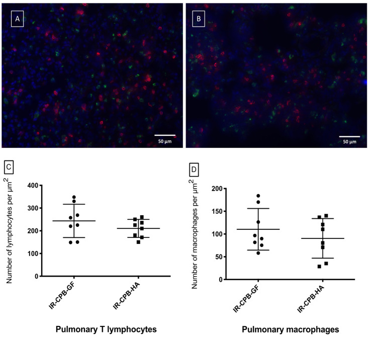
Figure 4.
Immunohistology (magnification x40, scale bar = 50 μm). Fluorescent immuno-histological marking of the pulmonary parenchyma of the IR-CPB-GF group (A) and the IR-CPB-HA group (B). For all the groups, macrophages (CD68) are marked in green and lymphocytes (CD3) are marked in red. (C) Pulmonary T lymphocytes (CD3) of the left lung tissue. (D) Pulmonary macrophages (CD68) of the left lung tissue. For statistical analyses, ANOVA was used, followed by Tukey’s multiple comparison (post hoc) testing. A result with p < 0.05 was considered to be statistically significant. Data are presented as mean ± SD. Abbreviations: CPB, cardiopulmonary bypass; IR, left lung ischemia–reperfusion; HA, human albumin.

