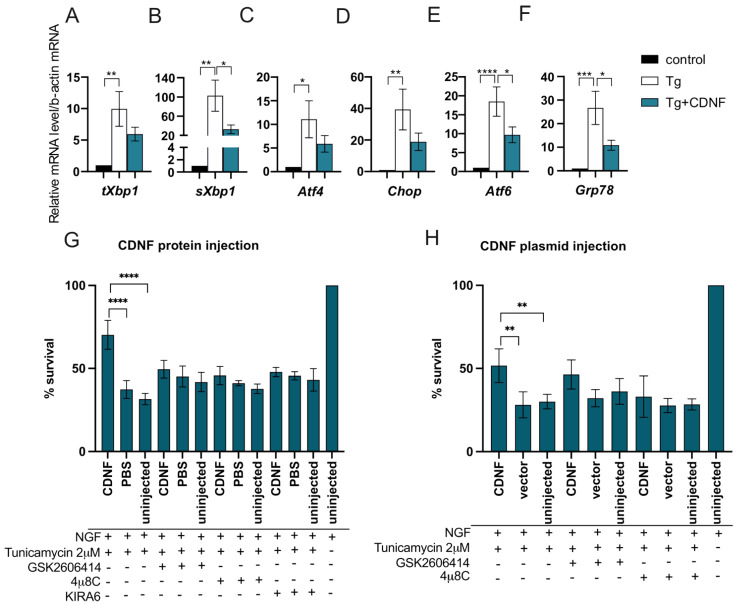
Figure 3.
CDNF promotes survival of ER–stressed cultured neurons and regulates UPR signaling in vitro. (A–F) CDNF treatment downregulated the expression of UPR genes sXbp1, Atf6, and Grp78 in embryonic mouse DA neurons under thapsigargin (Tg)-induced ER stress. DA neurons were cultured 5–7 days before adding CDNF (100 ng/mL) and inducing ER stress by adding 200 nM Tg. RNA was isolated from these neurons after 24 h. The expression levels of UPR marker transcripts were determined by qPCR. Each transcript’s levels have been normalized to beta-actin housekeeping gene and presented as a fold change to respective control data set from non-treated neurons. Shown are means of n = 10–18 experiments ± S.E.M. Repeated-measures ANOVA and Sidak’s multiple comparison post hoc test. (G,H) Survival-promoting activity of CDNF against ER stress is dependent on active IRE1α and PERK pathways. Mouse SCG neurons were microinjected with (G) recombinant human CDNF protein or (H) CDNF expression plasmid and treated with 2 µM tunicamycin and 2 µM PERK signaling inhibitor GSK2606414 or 25 µM IRE1α signaling inhibitor 4μ8C or 2 µM IRE1α inhibitor KIRA6. Then, 72 hours later, the number of living injected, fluorescent neurons was counted and expressed as the percentage of initially injected neurons. Shown are the means of 3–6 experiments ± S.D. Survival percentages in CDNF plasmid- or protein-injected groups were compared to the empty vector or PBS-injected controls of the same treatment group using ordinary one-way analysis of variance (ANOVA) and Sidak’s multiple comparison post hoc test. *, **, ***, **** denote p < 0.05, p < 0.01, p < 0.001, p < 0.0001, respectively.

