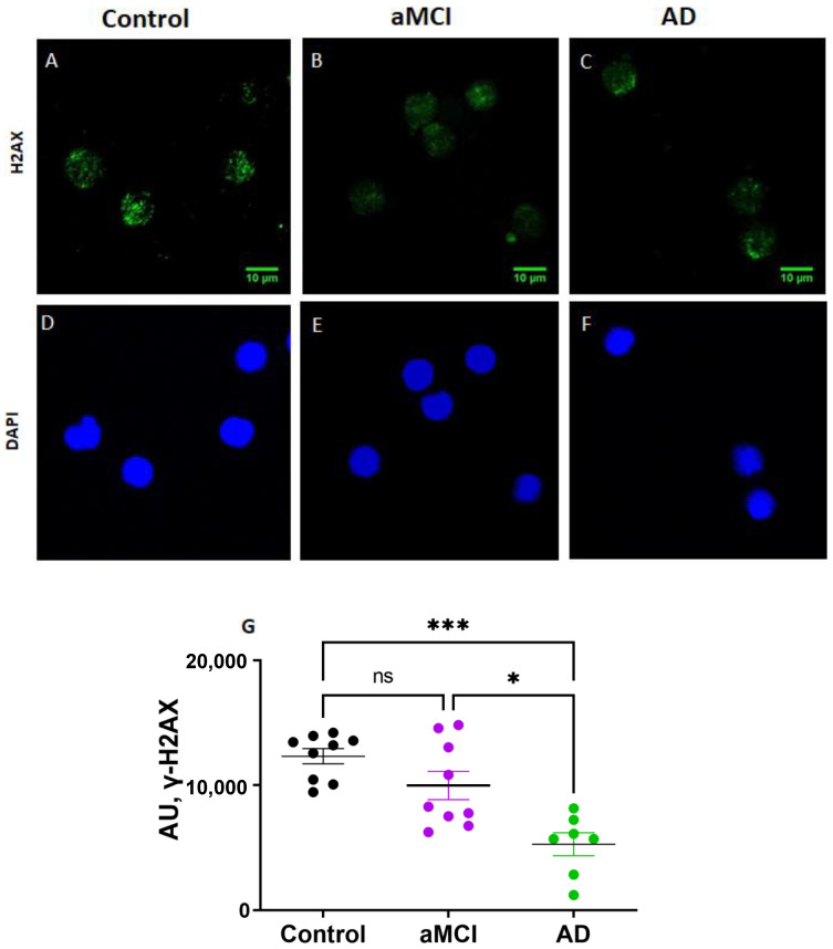
Figure 4.
Activation of the DNA damage response determined by γH2AX activity was decreased in PBMCs of AD patients. (A–F): Representative immunofluorescence confocal images of γH2AX staining (green, (A–C)) and nuclei stained with DAPI (blue, (D–F)) of PBMCs of a control (A,D), an aMCI (B,E) and an AD (C,F) patient. Scale bars: 10 μm. (G): Immunofluorescence quantification of γH2AX expression normalized by the number of nuclei in nine healthy controls, nine aMCI and seven AD patients. AU: arbitrary fluorescence units. Statistical analysis: Kruskal–Wallis with Dunn’s post hoc correction, * p < 0.05, *** p < 0.005, ns = non-significant.

