Figure 1.
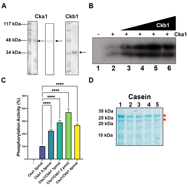
Purification and activity of fission yeast Cka1 and Ckb1. (A) Silver-stained PAGE-SDS and a Western blot (Cka1) of the purified subunits Cka1 and Ckb1. A 12% gel was used to separate the proteins and 200 ng of each subunit was analyzed. (B) Casein phosphorylation was determined using 2 pmol of Cka1 and increasing amounts of Ckb1 from 0.25 to 4 pmol (lanes 2–6)—indicates assay without Cka1 (lane 1). (C) The signal from each lane from (B) was quantified and plotted using the Image J software as described in Materials and Methods; – indicates a negative control without Cka1 and + indicates the assays with the addition of the enzyme Cka1. (D) A 14% PAGE-SDS gel was used to analyze the casein substrate used in (B) and shows that all lanes contain the same amount of substrate. Red asterisks show the phosphorylated polypeptides indicated in (B); **** indicates p < 0.0001.

