Figure 3.
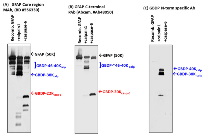
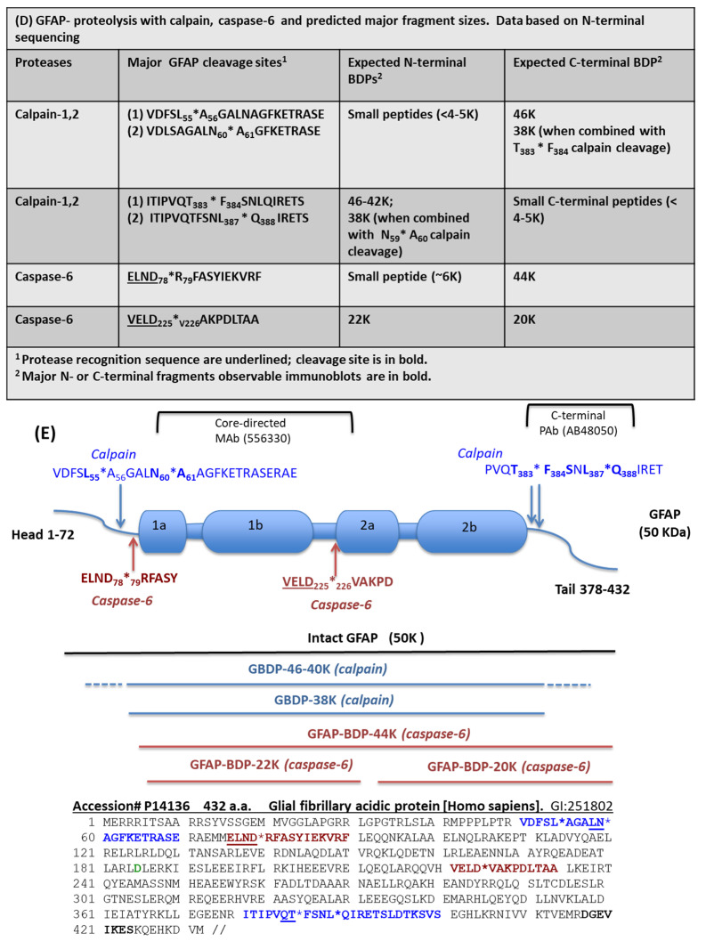
Digestion of purified GFAP by calpain-1 and caspase-6. Characterization with different GFAP antibodies and GBDP-specific antibodies by immunoblotting. (A) GFAP core Mab, (B) GFAP C-terminal Pab, (C) GBDP-specific antibody. (D) GFAP proteolysis with calpain, caspase-6, and predicted major fragment sizes. Data based on N-terminal sequencing. (E) Schematics of GFAP proteolytic fragment formation. The top panel shows GFAP linear model and major calpain/caspase-6 cleavage sites. The middle panel shows the positions of major fragments. The bottom panel shows the human GFAP amino acid sequence and the annotated cleavage sites (*) by calpain (blue), and by caspase-6 (red).


