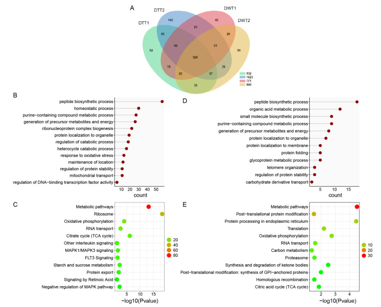
Figure 1.
The proteome analysis of DWT vs. DTT. (A) Venn diagram showing overlaps of protein in DWT and DTT. (B) Gene ontology enrichment analysis based on biological process (GOBP) of proteins that were downregulated in the presence of Wolbachia. (C) KOBAS pathway analysis of proteins that were downregulated due to Wolbachia infection. (D) GOBP analysis of proteins that were upregulated in the presence of Wolbachia. (E) KOBAS pathway analysis of proteins that were upregulated due to Wolbachia infection. DWT: Drosophila (Wolbachia-infected) testes; DTT: Drosophila (treated with tetracycline, Wolbachia-free) testes. (C,E): The color indicates the difference in protein counts. The redder the color, the more the count; the greener the color, the less the count.

