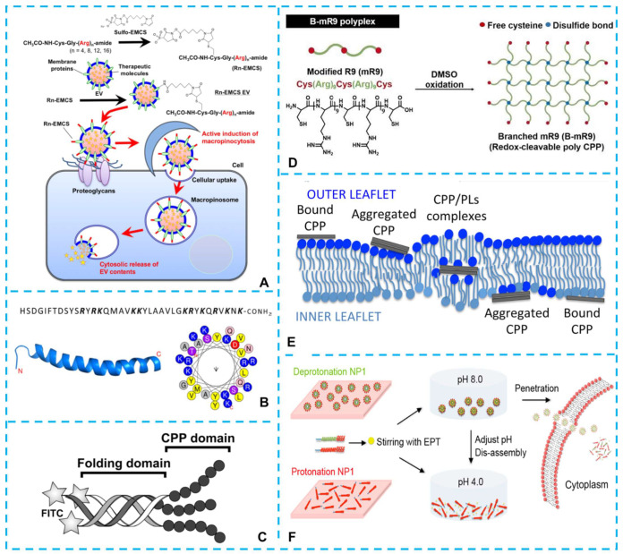
Figure 2.
(A) Oligoarginine peptides with different numbers of arginine residues (Rn: n = 4, 8, 12, 16) were used to modify the extracellular vesicles [60]. (B) Sequence and schematic ribbon representation of PACAP secondary structure and helical wheel representation of the putative a-helix segment of PACAP [87]. (C) Schematic representation of the CPP folded into a triple helix within the (POG)n sequence (folding domain). The CPP domain R6 or (RRG)2 was located at the C-terminus [89]. (D) Schematic illustration of the synthesis of the branched-modified R9 (B-mR9) CPP [91]. (E) Model of CPP direct translocation via the formation of inverted micelles. In this model, CPPs were internalized as neutral and hydrophobic complexes with anionic phospholipids (PLs) [CPPp+(PL-)p] [95]. (F) Schematic diagram of the pH-triggered self-assembly/disassembly of peptide NP1 to encapsulate and deliver the cancer drug ellipticine into cancer cells [36].

