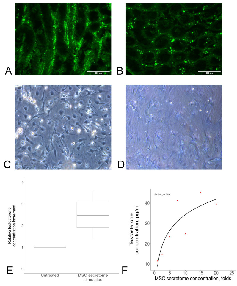
Figure 1.
(A,B) The locally injected mock product was predominantly distributed outside seminiferous tubules: (A) collagen (control); (B) collagen + GFP. (C,D) Morphology of Leydig cells: (C) 24 h after isolation; (D) 96 h after isolation. (E) MSC secretome-stimulated Leydig cells secreted more testosterone compared to untreated cells (the value of their potency was equal to one). Data are presented as the median, 25th and 75th percentiles, and minimum and maximum values. (F) Testosterone secretion by Leydig cells was directly associated with the MSC secretome concentration; the correlation was calculated using Spearman’s test (R = 0.82, p = 0.034). Red dots indicate independent samples.

