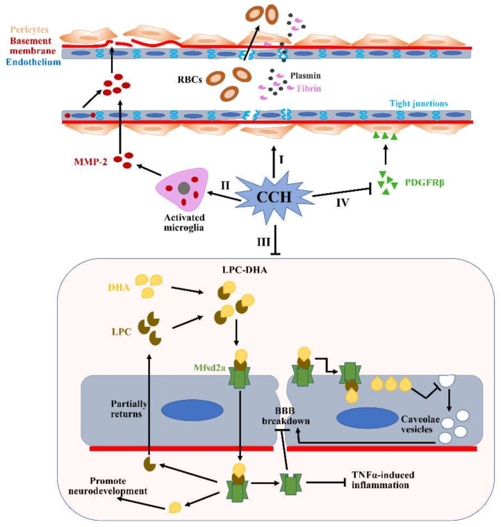
Figure 4.
Potential mechanisms of BBB breakdown during CCH. (I) Pericytes detach from the perivascular positions under the CCH condition, which subsequently reduce the expression of tight junction proteins. Additionally, CCH can also directly disrupt the expressions of tight junction proteins. (II) CCH can upregulate levels of MMP2 expression in the activated microglia and endothelium. Increased MMP2 can accelerate the degradation of many major ECM constituents, such as collagen IV, fibronectin and gelatin. (III) Mfsd2a plays a critical role in transporting DHA into the cells and maintaining the low rate of vesicular transcytosis. However, the expression of the Mfsd2a protein is significantly reduced during CCH, which causes augmented vesicular transcytosis. (IV) CCH-mediated PDGFRβ reduction can result in significantly decreased microvessel lengths. All of these pathological changes induced by CCH can subsequently lead to significant BBB impairment and contribute to the translation of many substances from the blood vessels to the brain parenchyma, such as RBCs, plasmin and fibrin. RBCs: red blood cells; DHA: docosahexaenoic acid; LPC: lysophosphatidylcholine.

