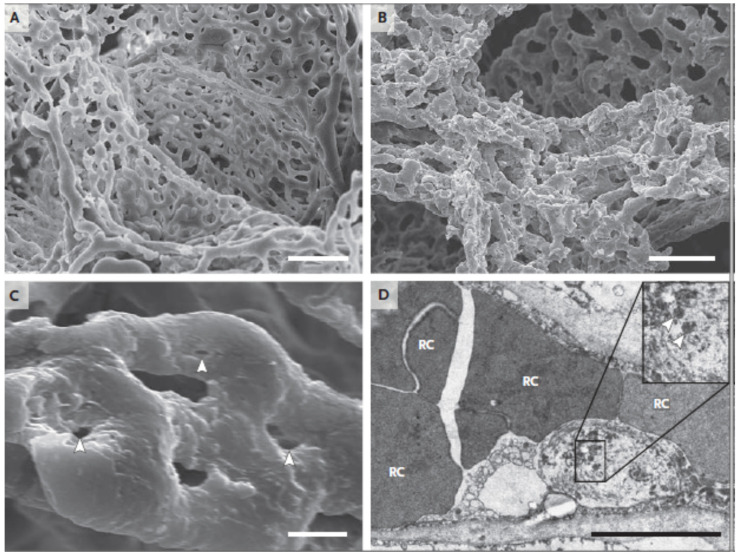
Figure 4.
Scanning electron micrographs of (A) microvascular corrosion casts from the thin-walled alveolar plexus of a healthy lung and (B) the considerable architectural deformation seen in lungs harmed by COVID-19. In (B), the disappearance of a vascular hierarchy that was visible in the alveolar plexus is attributed to the development of new blood vessels via intussusceptive angiogenesis. (C) The intussusceptive pillar localizations at higher magnification, indicated by the arrowheads. (D) Transmission electron micrograph demonstrating ultrastructural aspects of the breakdown of endothelial cells and the presence of SARS-CoV-2 within the cell membrane (arrowheads). The scale bar corresponds to 5 micrometers. RC stands for red cells [77].

