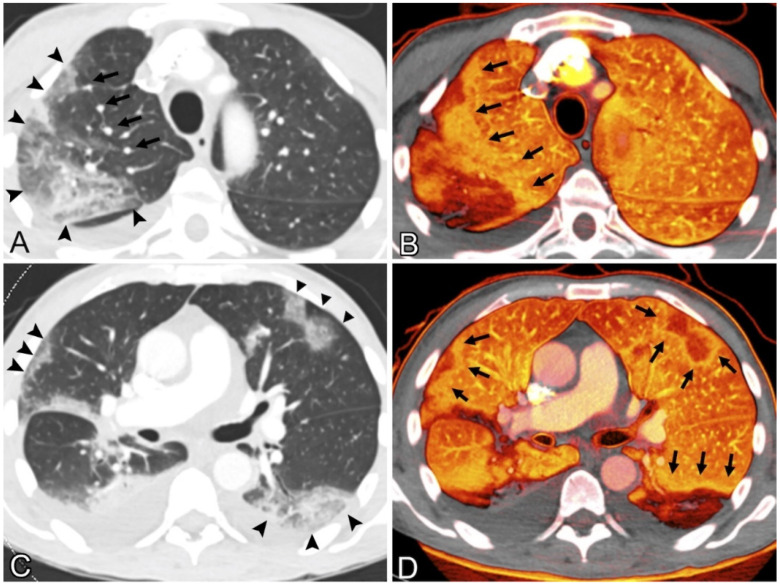
Figure 6.
(A) 69-year-old man with fever, weakness, and chills had coronavirus illness. The patient was hospitalized for acute intermittent tachycardia, desaturation, and shortness of breath. No pulmonary emboli were found. Contrast-enhanced CT pulmonary angiography of the upper lungs at lung windows showed ground-glass opacity and consolidation in the right upper lobe (arrowheads); sub-segmental arteries within the opacities were dilated, and right upper lobe vessels proximal to the opacity were similarly dilated (arrows). (B) Pulmonary blood volume (PBV) imaging at the same level shows a significant peripheral perfusion deficiency with a surrounding halo of enhanced perfusion (arrows). Heterogeneous left upper lobe perfusion. CT scan of the patient’s lower lungs showed peripheral ground-glass opacities and consolidation with a round or wedge-shaped appearance (arrowheads). (D) PBV picture shows perfusion deficiencies matching the opacities in (C), shown with enlarged perfusion halos (arrows) [82] (2020).

