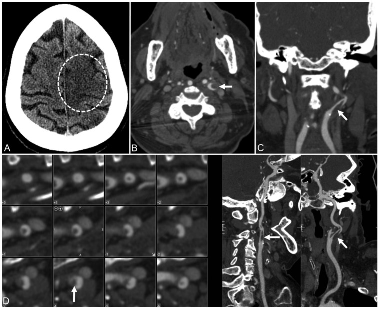
Figure 12.
Patient 2. (A) 78-year-old woman with COVID-19 and an NIHSS score of 25.The CT of the head without comparison shows an evolving ischemic infarct in the left frontal brain paracentral cortex (dotted circle) and a smaller infarct in the left parietal cortex. (B–D), Axial, coronal, and curved reimaged images from CT angiography of the head and neck show an irregular plaque at the left internal carotid artery bifurcation and a capillary filling defect (arrow) extending superiorly in the left internal carotid artery, which matches the ruptured plaque with clot formation [144].

