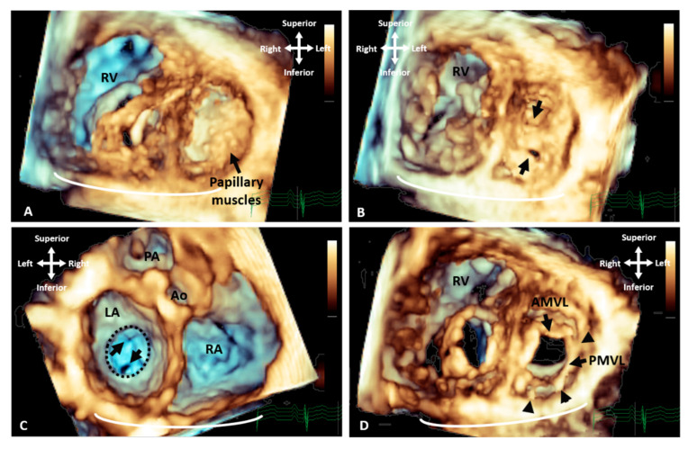
Figure 2.
Three-dimensional TTE demonstrating the appearance of parachute mitral valve with complex subvalvar stenosis. (A) Demonstrates the thickened and fused subvalvar apparatus with indistinguishable papillary muscles and chords as seen from a distal ventricular view. A view through the middle of subvalvar tissue reveals two small orifices as seen from the (B) ventricular and (C) atrial aspects, the atrial view also shows the small annulus that is highlighted with the dashed line. (D) A view at the level of the mitral valve leaflets shows that although the posterior leaflet is tethered with multiple short chords (arrowheads), the anterior leaflet retains adequate excursion, with area of narrowing and stenosis being within the subvalvar tissue. Ao: Aorta; AMVL: Anterior mitral valve leaflet; LA: Left atrium; PA: Pulmonary valve; PMVL: Posterior mitral valve leaflet and RA: Right atrium.

